Scans
Brain Scans
When a person develops dementia, their brain loses brain cells, and specific patterns in the brain will indicate this loss. A radiologist viewing the scan looks out for particular signs of damage, for example shrinking of the hippocampus and presence of amyloid plaques.
Brain imaging techniques are available to define the structure, function, composition and molecular pathology of the brain. These techniques can be classified as either structural or functional, depending on the type of information that they provide.
Structural: CT (computed topography) and CAT (computerized axial tomography) use X-rays and a computer to provide structural information. A more sensitive technique is MRI scanning.
MRI scans
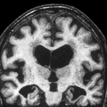
Magnetic resonance imaging (MRI) scans give high quality images that reveal physical changes in the brain. MRI scans also create a computer image of the brain from radio signals produced by the body in response to the effects of a very strong magnet.
 It is well established that techniques like structural MRI can be used to detect changes in brain structure that are thought to become evident later during the disease progression and to match more closely the cognitive symptoms26.
However, it is now becoming apparent that techniques like MRI can also contribute to a better understanding of the sequence of events occurring before Alzheimer’s manifests clinically. Scientists have pioneered the development of a computer program to interpret the results of MRI scans in more detail than the human eye and plan to test the potential of this technology in diagnosing dementia at an earlier stage than is currently possible33.
For further information defining CT and MRI scans please follow this link:
Click here to go to the www.brainandspine.org site »
Functional: PET (positron emission tomography) and SPECT (single photon emission computerized tomography) scans look at the blood flow through the brain, and metabolic activity, rather than at the structure of the brain.
PET and SPECT are used for evaluating cognitive impairment and distinguishing among primary neurodegenerative disorders22.
Positron emission tomography (PET) scan
A PET (positron emission tomography) scan reveals chemical functions rather than physical structures. A small dose of radiation is introduced into the body before a PET scan using a medicine called a radiotracer. A PET scan works by detecting this radioactive ‘tracer’, which concentrates in a specific part of the body.
PET scans are useful for early diagnosis because they can identify changes to brain metabolism that may occur in Alzheimer’s disease before structural changes occur.
Learn more about Biomarkers »


